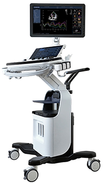
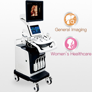
System Overview
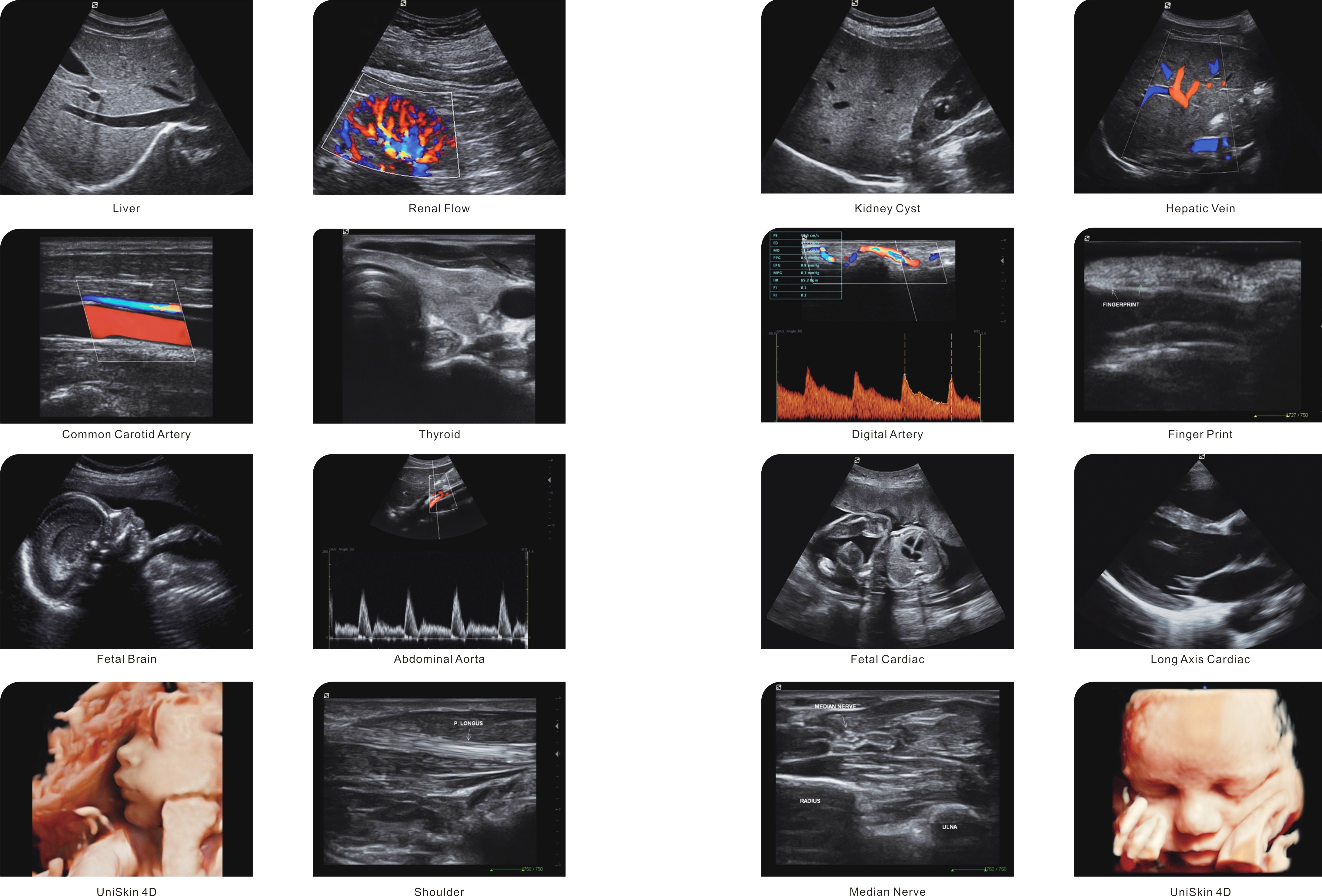
Ứng dụng
Abdomen-Bụng
Obstetrics-Sản
Gynecology-Phụ khoa
Cardiology-Tim
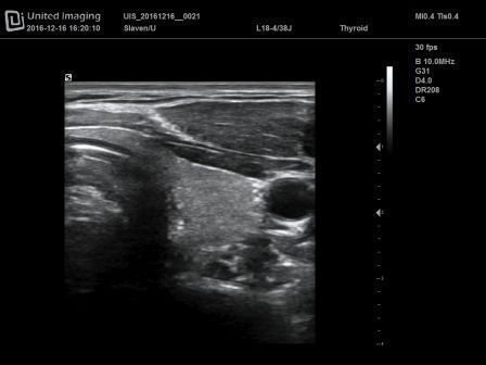
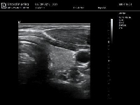
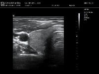
Small parts-Các phần nhỏ
Urology- Tiết niệu
Vascular-Mạch máu
Pediatrics-Thai nhi
Nerve-Thần kinh
Others- Các phần khác
Các loại đầu dò
Đầu dò cong
Đầu dò linear
Đầu dò tim
Đầu dò 4D
Các chế độ hình ảnh
B-Mode –Chế độ B
(Tissue Harmonic Imaging & Pulse Inversion Harmonic Imaging-Hình ảnh hài hòa mô và hình ảnh hài hòa mạch)
M-Mode-Chế độ M
Anatomical M-mode-Chế độ M cắt lớp
PSI (Patient Specific Imaging)-Hình ảnh bệnh nhân đặc thù
TSI (Tissue Specific Imaging)
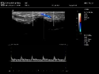
Color Doppler Imaging-Hình ảnh Doppler màu
Power Doppler /Directional PD- Doppler năng lượng/ PD trực tiếp
Pulsed Wave Doppler-Doppler sóng mạch
Continuous Wave Doppler-Sóng doppler liên tiếp
4D
UniLiveTM 4D
Standard features-Cấu hình chuẩn
B-Mode –Chế độ B
(THI & PHI)
M-Mode
Anatomical M-mode
PSI (Patient Specific Imaging)
TSI (Tissue Specific Imaging)
Color Doppler Imaging
Power Doppler and Directional PD
Pulsed Wave Doppler
Continuous Wave Doppler
Doppler auto calculation & parameter-Tham số và tự động tính toán Doppler
Duplex-Kép
Triplex-
Dual Screen-Màn hình kép
Quad Screen- Màn hình bốn
UniCTTM Hình ảnh phức hợp không gian
UniViewTM (Adaptive Speckle Reduction)
UniFCITM (Frequency Compound Imaging)
iuScanTM (Auto Optimization)
Auto OB (BPD, HC)
Zoom
B steer
Trapezoid
Login User section:
Set admin/user/guest login password;
Set user permissions;
4 active probe ports
1TB hard drive
6 USB ports (2 located in the front of the system for user convenience)
Optional features
4D
UniLiveTM 4D
Continuous Wave Doppler
DICOM
ECG
Biopsy Guide
Video & PC printer
WIFI access (Able to connect WIFI, wireless printer printing, WIFI DICOM transfer)
UniTutorTM (Anatomical diagram illustrations comparison with real-time scanning demonstrating patient position and probe placement tips)
Specialized breast screening program
Physical Specification
Dimension and weight
Height: 1512mm
Width: 565mm
Depth: 851mm
Trọng lượng: Khoảng 85kg riêng máy)
Monitor-Màn hình
Màn hình LED độ phân giải cao 19inch
Điều chỉnh độ sáng độ tương phản trên màn hình
Độ phân giải: 1280 ×1024
Audio speakers-Loa
Loa âm thanh nổi
Màn hình xoay đa chiều thuận tiện cho người dùng
Xoay: ±90 độ (R/L, F/R)
Lên/xuống: ±30 độ
Wheels
Đường kính: 125mm
Bánh tránh (2 cái): Khóa và phanh
Bánh sau (2 cái) Khóa và phanh
Probe port and holder-Cổng và giá đỡ đầu dò
Cổng cắm đầu dò: 4
Gía để đầu dò: 5 thêm 1 giá để đặc biệt đầu dò âm đạo
Electrical Power-Điện thế
Điện thế: 110V (± 10%) ~ / 220V (± 10%) ~
Tần số: 50/60 Hz
Lượng điện tiêu thụ: Approx. 300VA
Bộ ngắt mạch: 110V:125V~, 10A; 220V: 250V~, 5A
Môi trường thao tác
Nhiệt độ phòng: 0 – 40 °C
Độ ẩm tương đối: 30% – 75% (no condensation)
Atmospheric pressure: 700hPa – 1060hPa
Storage & Transportation Environment
Ambient temperature: -20 – 60 °C
Relative humidity: 20% – 90% (no condensation)
Atmospheric pressure: 700hPa – 1060hPa
User Interface
Control panel
User-centric control panel
Backlit keys ensure accurate work in the dark room
Programmable keys available for user-defined functions
8-segment TGC control
Full-sized, wireless, QWERTY keyboard for text input, function keys and system programming
Dedicated palm rest design to help reduce user repetitive stress injury
Independent rotation and up/down of control panel facilitates optimal positioning
Rotate: ±90 degrees (from center) (optional)
Down/up: 100mm
Touch screen
11inch high sensitivity anti-glare color touch screen
On-screen display of brightness and contrast controls
Support with gloves on
Viewing angle: ≥150 degrees
Resolution: 800 x 600
System boot-up
Boot-up from complete shut-down in less than 100 sec
Shut-down in less than 30 sec
Comments
Supports text input and arrow
Adjustable text size
Covers various application
User customizable
Body mark
More than 110 body marks for versatile application
User customizable
Screen information
Common info:
United Imaging logo
Hospital name,
Exam date
Exam time
Thermal and Mechanical Acoustic Output Indices
Gender, Age, ID, Last name, First Name, Middle Name
Probe model
TGC Curve
Focus position
Thumbnail
Imaging parameters
Imaging Parameters
Overview
Hybird beam-forming
280 dB full time input dynamic range
65536 digitally-processed channels
Parallel RF signal processing, quad beam-forming
Image depth up to 30 cm (exam and transducer specific)
B-mode
Depth
Zoom Real time, Freeze
Frame rate Up to 640 fps
TGC 8-segment TGC control
Dynamic range 280 dB full-time input dynamic range Noise rejection
Gain
Persistence 5 levels (exam and transducer specific)
L/R flip and U/D flip
Gray Map 8 levels (exam and transducer specific)
Frequency
Chroma map
Acoustic Output Indices
UniviewTM: Adaptive Speckle Reduction
iuScanTM: Auto Optimization
UniFCITM: Frequency compound imaging
UniCTTM: Spatial compound imaging
B steer: Linear probe only
FOV: FOV available on all transducers
Focus number: Adjustable
Focus position: Adjustable
Focus spacing: Adjustable
Line density: 64/128/256
Display formats: Single (B), Dual (B+B), Quad (4B)
TSI: General/Fat/Fluid/Muscle
Pulse Inversion THI
Available on all types of transducer
Utilizing pulse inversion technologies to enhance high-resolution harmonic signals, iuStar300 provides increased contrast and axial resolution, clearer boundaries and better visualization of structures
M-mode
Display formats: V1:1, V1:2, H1:1, FULL (V: vertical, H: horizontal)
Dynamic range
Gain
M sweep speed
Chroma map
Gray Map
Time mark
L/R flip and U/D flip
Acoustic Output Indices
Color Doppler Imaging
ROI size/position: Adjustable
Display formats: Single (B + Color), Dual (B + Color), Quad (B + Color)
Color Doppler velocity: ±2.1 – ±308 cm/sec (with baseline correction)
Scale.E: 0/1/2
B/C Compare: On/Off (Side by side displays B and B + Color)
Line density: L/H
Steer (linear probe only)
Gain
Scale
Baseline
Frequency Adjustable, independent of B-mode
Wall Filter
Packet size
L/R flip and U/D flip
Color map
Invert: On/Off
Persistence
Acoustic Output Indices
Power Doppler Imaging
ROI size/position: Adjustable
B/C Compare: On/Off (Side by side displays B and B+PD)
Line density: L/H
Support directional power Doppler
Gain
Steer (linear only)
Scale
Scale.E
Wall filter
Packet size
Color map
Frequency Adjustable, independent of B-mode
Directional color map
Persistence
L/R flip and U/D flip
Acoustic Output Indices
PW/CW Mode
Định dạng hiển thị: V1:1, V1:2, H1:1, FULL
(V: Dọc H: ngang)
Duplex/Triplex (PW only)
PW vận tốc: ±0.8– ±385 cm/s với góc chỉnh 0°
vận tốc CW: ±1.9 – ±739 cm/s với góc chỉnh 0°
Kích thước khối lượng mẫu (SV) (PW only)
Vị trí cổng mẫu: có thể chỉnh
Thang .E (chỉ trong PW )
Steer (chỉ đầu dò linear)
Tham số tính : PS, ED, MD, TAMAX, PPG, EPG, MPG, VTI, AT, DT, Time, HR, PI, RI, S/D, D/S
Diện tích trace: Top, Bottom, Both.
Scale
Baseline
Volume
Frequency Adjustable
Gain
Dynamic range
Sweep speed
Wall filter
Invert
góc PW chỉnh góc 0-80°
(+60°, 0°,-60° and 2° increments)
góc.E
Gray map-bản đồ xám
Chroma map- bản đồ màu
Auto calculation – tự động tính
Chỉ số đầu ra âm thanh
Static 3D and 4D
Rotation: 0º, 90º, 180º, 270º, X, Y, Z
Render modes: Surface, Min, Max, X -ray
Active quadrant: A, B, C, 3D
Display formats: Single, Dual, Quad
Render modes: Surface, Min, Max, X -ray
Smooth: off, E1, E2, E3
Inversion: ROI/UD ROI/LR
Line Density: Low, Mid, High
Image quality: Low2, Low1, Mid, High1, High2
Zoom: off, L1, L2, L3, L4, L5
Edit: Polygon, Ellipse, Rect, Erase
Angle
Mouse ROI
Panning
Threshold:
Brightness
Contrast
Colorize Map
Slice
Save
Thumbnail Image Display
Support Save, Send to USB, Disc writer, Printer, Network
Image/cine review
Available in all modes
Cineloop image review (up to 3000 B/W frames)
Frame by frame manual cine loop review or auto playback with variable speed
Retrospective and prospective storage are available and length is pre-settable
B-mode review up to 266 sec for retrospective analysis
Doppler review up to 40 sec for retrospective analysis
Jump to first and jump to last: one stroke go to first or last frame in the cine
Raw data processing:
B-mode: Gain, Dynamic range, Gray map, Chroma map, flip, UniViewTM, TGC
Color: Gain, Baseline, Invert, Color map
M-mode: Gain, Gray map, Chroma map
Doppler: Gain, Dynamic range, Gray map, Chroma map, Baseline, Invert, Angle, Angle.E, Auto Calculate, VMax, VMean
Measurement, Comment
4D:ROI, slice, Threshold, Contrast, Brightness, X, Y, Z, UniLive
Quick save (Able to save image during real-time scanning without freeze)
Measurement/Analysis and Report
Generic measurements
2D-mode
Depth
Distance
Angle
Ratio (D)
Trace Length
Area: Ellipse, Trace
Volume: 3-Distance, Ellipse, Ellipse + Distance
M-mode
Distance
Time
Slope
Heart Rate
Depth
Doppler mode
D Velocity
Time
Heart Rate
Acceleration
PS/ED
Ratio (V)
Clinical option measurement package
Abdomen
B/C mode
Flow Diam., Liver, CHD, CBD, Portal V Diam., Spleen, Aorta Diam., Iliac Diam.
Gallbladder: GBL, GBH, GB WALL TH
Pancreas: head body tail duct
Renal: Renal L, Renal H, Renal W, Cortex, Renal vol.
Doppler mode:
PI, D Trace, M Renal A, Segment A, Interlobar A, Arcuate A, Renal V, Aorta, Celiac Axis, SMA, Hepatic A, Splenic A,
Abdom venous: IVC, Portal V, Hepatic V, M Hepatic V
Splenic V, SMV
Gynecology
B/C mode:
Uterus: Length, Height, Width, UT Vol, Endo Thickn
Uterine Cervix: Cervix L, Cervix H, Cervix W
Ovary: Length, Height, Width, Ovary Vol
Follicle1 Follicle2 Follicle3: L, W, H, Vol
Vol Flow: Vas Diam
Doppler mode:
D Trace, Ut A, Ovarian A
Vol Flow: Vas Diam, Trace.Manual, Trace.Auto
Obstetric
B/C mode:
Fetal Biometry: BPD, HC.Ellipse, HC.Trace, AC.Ellipse, AC.Trace. FL, HL, APAD, TAD, APTD, TTD, FTA.Ellipse, Cereb, OFD, MAD, FTA.Trace
Early Gest: CRL, GS, YS, BPD, FL, NT (Semi-auto NT: Nuchal Translucency Measurement)
Long Bones: HL, RAD, Ulna, TIB, FIB, Clav
Fetal Cranium: Cereb, CM, BOD, IOD
AFI: AF1, AF2, AF3, AF4
Uterus: Length, Height, Width, UT-Trace, Endo Length, Endo Thickn, Cervix L , Cervix H
Ovary: Length, Height, Width
Umbilical Vein: Diam
Doppler mode:
Ductus Art, Ao, Carotid, MCA, Umb A, Ductus Vein, UT A, FHR, SMA, Celiac A.
Umbilical Vein: TAmax
Kidney
B/C mode:
Renal: Renal L Renal, H , Renal W, Cortex, Renal vol.
Doppler mode:
PI, M Renal A, Segment A, Interlobar A, Arcuate A, Renal V, Aorta, Celiac Axis, SMA, Hepatic A, Splenic A,
Abdom venous: IVC, Portal V, Hepatic V, M Hepatic V, Splenic V
Urology
B/C mode:
Flow Diam , Vol Flow
Renal: Renal L, Renal H, Renal W, Cortex, Renal vol
Prostate: Prostate W, Prostate H, Prostate L, Prostate Vol\
Seminal vesicle: Seminal L, Seminal H, Seminal W
Bladder: Pre-BL L, Pre-BL W, Pre-BL H, Post-BL L, Post-BL W, Post-BL H
Doppler mode:
D Trace, Vol Flow, M Renal, A, Segment A, Interlobar A, Arcuate A, Renal V
Cardiac
B mode:
LA Diam, AO Diam, IVSd, LVPWd, RVOT Diam, RVDd, EPSS, LVOT Diam, IVC Diam(Insp), IVC Diam(Exp), SVC Diam(Insp), SVC Diam(Exp), VSD Diam, ASD Diam
Aortic Valve: ACS, AVA(Ellipse), AVA(Trace)
MV : MV Diam, MCS, MVA.Ellipse, MVA.Trace
Simpson: AP4d, AP2d, AP4s, AP2s
AL: Ad, Ld, As, Ls
Pombe: LVIDd, LVIDs
PA: MPA Diam, LPA Diam, RPA Diam
Left Atrium: LA Anteroposterior, LA Major, LA Minor, LA Area.Ellipse, LA Area.Trace
Right Atrium: RA major, RA Minor, RA Area.Ellipse,
RA Area.Trace
Ventricle: Ven Major, Ven Minor, Ven Area(d).Ellipse, Ven Area(d).Trace, Ven Area(s).Ellipse, Ven Area(s).Trace
AORTA: AortaRings, Ao Sinus Diam, Ao ST Junction, Ao Asc Diam, Ao Arch Diam, Ao Desc Diam, Ao Isthmus, Duct Art Diam
TVA: Ellipse, Trace
M mode:
LA Diam, Ao Diam, EPSS, Cardic Teich, Cardic Pombo, ACS, IVSd, LVPWd, RVOT Diam, RVDd, ,LVPEP, LVET, RVPEP, RVET, Cardic Gibson, Cardic Teich.E
MV: MV DE, MV E-F slope, EPSS, MV E Amp, MV A Amp
Doppler Mode:
MV: MV E Vel, MV A Vel, MV DecT, MV A Dur
MV VIMP: IVRT, IVCT, ET
LVOT VTI: LVOT Diam, LVOT VTI.Manual, LVOT VTI.Auto
MV SV: MV Diam, MV.Manual, MV.Auto
Aortic Valve: AV Vmax, LVOT Vmax, LVET
Pulmonic: RVOT Vmax, PV AccT, RVET
RVOT VTI,
Pulmonary Vein: Pvein S Vel, Prein D Vel, Pvein A Vel, PVein A Vdur.
Tricuspid Valve: TV Vmax, TV E Vel, TV DecT, TV A Vel
TV VIMP: TV IVCT, TV IVRT, TV ET.
TEI: ICT, IRT, ET.
Vascular
B./C mode:
Stenosis (A): Ellipse, Trace
Stenosis (D)
Flow Diam
Vol Flow
Semi-auto IMT (Intima-Media Thickness measurement)
Doppler mode:
Carotid:

 Call for me!